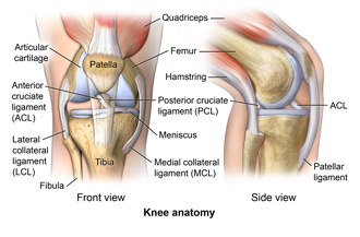
Anatomy of the Knee
 |
| Click image to enlarge |
The knee is a vulnerable joint. It bears a lot of stress from everyday activities, such as lifting and kneeling. It also bears stress from high-impact activities, such as jogging and aerobics.
The knee is formed by the following parts:
-
Tibia. Shin bone or larger bone of the lower leg.
-
Fibula. Smaller of the 2 lower leg bones.
-
Femur. Thighbone or upper leg bone.
-
Patella. Kneecap.
Each bone end is covered with a layer of cartilage that absorbs shock and protects the knee. Basically, the knee is 2 long leg bones held together by muscles, ligaments, and tendons.
There are 2 groups of muscles involved in the knee:
Tendons are tough cords of tissue that connect muscles to bones. Ligaments are elastic bands of tissue that connect bone to bone. Some ligaments on the knee provide stability and protection of the joints. Other ligaments limit forward and backward movement of the tibia or shin bone.
Online Medical Reviewer:
Rahul Banerjee MD
Online Medical Reviewer:
Raymond Turley Jr PA-C
Online Medical Reviewer:
Stacey Wojcik MBA BSN RN
Date Last Reviewed:
1/1/2023
© 2000-2025 The StayWell Company, LLC. All rights reserved. This information is not intended as a substitute for professional medical care. Always follow your healthcare professional's instructions.